Ph.D. Thesis
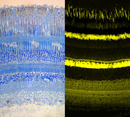
Several species of anchovies (Engraulidinae) feature unique structure and arrangement of photoreceptors amongst vertebrates: the position of photopigment containing lamellae, i.e. light-parallel and perpendicular to each other in two cone types, allows for the perception of linearly polarized light (two-channel-analyzer). So far, only the outer retina of anchovies, including photoreceptors, pigment epithelium and tapetum, is described in detail (Fineran & Nicol, 1976, 1977, 1978; Zueva, 1980; Heß, 2000, 2009; Heß et al., 2002, 2006).
In the present study, a multi-methodical approach to study the inner retina was accomplished to finally obtain a comprehensive overview of morphology, ultrastructure, connectivity and density distribution of neuronal elements in the specialized retina of the European anchovy Engraulis encrasicolus L.
The histology of the inner retina was analyzed light-microscopically on the basis of semi-thin and fluorescently labeled sections. Thereby, particular attention was paid to the analysis of the inner plexiform layer by proposing a possible sublayering.
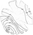
A first picture of cell density distributions, cell convergences and divergences is provided by the topography of different cell types, classified by fluorescently labeled nuclei and evaluated quantitatively. The cell density maps of the inner retina show a clear correlation between most neuronal elements and the cone density distribution (with maxima in the ventro-temporal quadrant and the dorso-nasal periphery), other cell correlations could be displayed with simple arithmetic operations.
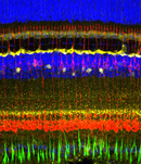
Immunohistochemistry succeeded in refining the cell classification and therefore the cell topography and could reveal the neuroanatomy of different cell populations. First findings about connectivity of secondary order neurons with photoreceptors were offered by this method.
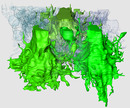
By electron microscopical analysis and 3-dimensional reconstruction of the neuronal network of the anchovy retina with the new method of FIB-FESEM (focused ion beam field emission scanning electron microscopy) a freely manipulable, high-resolution 3D-model of all neuronal elements in the volume of interest (VOI) could be generated, displaying detailed morphologies and connectivities of all retinal neurons and Müller cells of the anchovy retina for the first time.The spatial and synaptic relations of three horizontal cell types, nine bipolar cell types and numerous amacrines and ganglion cells were described in detail: the dendrites of horizontal cells 1 (H1), showing a synaptic preference for long cones but contacting short cones as well, embrace the synaptic ribbons of photoreceptors laterally (like all horizontal cell types), whereas up to three different h-cells contact the same synaptic ribbon. H2 exclusively contact short cones, H3 are synaptically linked to rods. We suggest, that the anchovy polycone is composed by the original red- and green-sensitive cones of teleosts, whereas the blue-sensitive cone is lost. The nine bipolar cell types can be classified in ON- and OFF-cells depending on their termi-nation depth in the IPL, whereas the shape of axon terminals is highly variable. Their different connectivity patterns show preferences for one to two photoreceptor types each. A detailed analysis for amacrines and ganglion cells is not yet possible, as their dendritic fields are too wide to be analyzed in that limited VOI using the method of FIB-FESEM.
The results of the different methodic approaches are discussed with respect to the primary literature. Finally, an attempt for a comprehensive and comparative methodical discussion is made. Previous structural data of the anchovy retina are refined and extended by the interaction of the methodical approaches presented here and provide extensive new findings about neuro-anatomy, ultrastructure and connectivity patterns in the retina of a vertebrate that is sensitive to polarization contrast.